Vestibular disorders
Semicircular canal dehiscence
This information is intended as a general introduction to the topic. Since every person is affected differently by balance and dizziness problems, you should consult your doctor for individual advice.
For reasons of readability, the generic masculine form is used and the simultaneous use of the male, female, and diverse (m/f/d) forms is omitted. Unless otherwise specified, the personal designations used in this patient information refer to all genders.
What is a semicircular canal dehiscence?
Semicircular canal dehiscence is a cluster of hearing and balance problems caused by a tiny hole (called a dehiscence) in one or more of the semicircular canals in the inner ear. Usually the superior semicircular canal is affected, but sometimes there is also a hole in the posterior semicircular canal. Dehiscence of the semicircular canals can affect one or both ears. It was first described in 1998.
It is not clear how many people suffer from semicircular canal dehiscence. It seems to be more common in older adults, but young children can also have semicircular canal dehiscence. Some people have a hole in the semicircular canal but show no symptoms.
It is not clear how many people suffer from semicircular canal dehiscence. It seems to be more common in older adults, but young children can also have semicircular canal dehiscence. Some people have a hole in the semicircular canal but show no symptoms.
Summary
- A rare disease, usually caused by a hole in one of the bony coverings of the semicircular canals in the inner ear.
- The superior semicircular canal is usually affected.
- It is most common in older adults, but can occur at any age.
- The hole can occur (A) during fetal development, (B) in connection with an injury, or (C) later in life as the bones become thinner.
- It can cause both hearing and balance problems.
- Patients often hear internal sounds such as their own voice, heartbeat or even the movement of the eyeball.
- Avoiding triggers, tinnitus retraining therapy and the use of a hearing aid can help to improve symptoms.
- If the symptoms are severe, surgery is suggested to close the hole.
What causes semicircular canal dehiscence?
The inner ear consists of two interconnected structures: the vestibular organ, which is needed for balance, and the cochlea, which enables hearing. These structures are surrounded by bone and filled with a fluid (endolymph).
There are three semicircular canals in each ear, which are arranged at right angles to each other. They perceive head inclinations and rotations. For example, when you nod your head, the sluggish endolymph in the posterior semicircular canal initially remains still while the semicircular canal moves. This causes the endolymph to press against special hair cells that signal to your brain that your head is moving.
The cochlea is a tube that is rolled up and looks like a snail shell. It converts sound waves into nerve signals that are transmitted to your brain so that you can hear. The sound waves enter the ear canal and cause the eardrum to vibrate, which is transmitted to the stapes bone. This tiny bone presses on the oval window. This structure is located at one end of the cochlea and causes pressure waves in the endolymph within the cochlea. The organ of Corti in the cochlea converts these pressure waves into nerve signals. Finally, the pressure waves leave the cochlea through another structure, the round window. The round window acts like a drain valve for the cochlea: it moves outwards when the oval window moves inwards, and vice versa.
The oval window and the round window are membranes. The rest of the inner ear is rigid because it is enclosed by bone. Normally, the round and oval windows are the only structures of the inner ear that move in response to the sound waves so that the sound waves stay in the cochlea. However, if there is a hole in the bone covering one of the semicircular canals, there is now a "third window" in the inner ear. This means that some of the sound waves do not remain in the cochlea, but can escape into the semicircular canal. This leads to several problems:
- The pressure of the sound waves in the cochlea is lower, so they cannot all be converted into nerve impulses. This means that you cannot hear some sounds well.
- The sound waves can press against the hair cells of the semicircular canals, causing your brain to think your head is moving when it is not.
- Sound waves from inside the body can enter the semicircular canals and then the cochlea, causing these internal body sounds to be distorted or louder than normal.
Sometimes the hole is up to two millimeters in size. However, the hole can also be as small as a pinhole and still cause symptoms.
The symptoms of semicircular canal dehiscence vary from person to person. Both hearing and balance disorders can be caused.
Balance disturbances in semicircular canal dehiscence are often caused by loud noises or pressure changes (either inside the body, e.g. when coughing, or outside the body, e.g. when taking off or landing an airplane). These can include
- Vertigo and nystagmus (rapid, involuntary eye movements) in response to loud noises (Tullio phenomenon) or changes in pressure (Hennebert's sign)
- Postural and gait instability (balance disorder)
- Oscillopsia, blurred or shaky vision in response to loud noises or pressure fluctuations
- A feeling of fullness in the ear
Possible hearing disorders of semicircular canal dehiscence:
- A form of hearing loss known as pseudo-conductive hearing loss in the low-frequency range. "Low-frequency" means that it is more difficult to hear sounds with a lower pitch. "Pseudo-conductive hearing loss" means that your middle ear function is normal, although some of the hearing test results resemble conductive hearing loss.
- You hear unusually loud noises from your own body (bone conduction hyperacusis), e.g. your voice (autophony), your heartbeat, the cracking of your joints or even the movement of your eyeballs
- Hearing rhythmic noises that seem to have no external source (pulsating tinnitus)
- Sounds from outside the body or your own voice are distorted or difficult to hear
- Sensitivity to loud noises (phonophobia), as they can cause the Tullio phenomenon
Diagnose der Bogengangsdehiszenz
Dehiscence of the semicircular canal is usually diagnosed by a neurologist or ENT specialist who specializes in dizziness.
These specialized doctors will take a detailed medical history, perform a neurological examination, and carry out various tests to assess the function of your vestibular system.
You will likely undergo the following diagnostic tests:
- Hearing and balance tests, including audiometry, vestibular evoked myogenic potentials (VEMP) and video nystagmography (VNG)
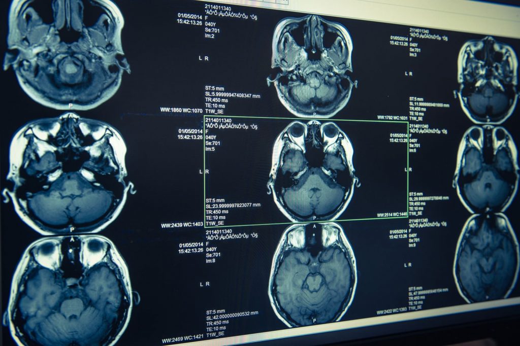
Imaging (high-resolution CT scan)
Treatment of semicircular canal dehiscence
Currently, the only treatment option is surgery. However, if your symptoms are mild, the disadvantages of surgery may outweigh the benefits. Your doctor may suggest you start with less invasive methods to treat your symptoms.
Some people find it helpful just to know what is causing their symptoms. Other people find that their symptoms improve with different techniques, e.g:
- Avoid triggers such as loud noises and music; earplugs can help to muffle sounds
- Avoid strong pressure changes, e.g. when diving or flying
- Avoid lifting or straining
- Avoid equalizing pressure in the ear or blowing your nose vigorously
- Tinnitus retraining therapy
- Use of a hearing aid
Your doctor will examine you regularly to see how you are doing and whether your symptoms are changing. Dehiscence of the semicircular canal will not get better on its own and your symptoms may stay the same or even get worse without surgery. Symptoms may increase with age as the thickness of the bone over the superior semicircular canal decreases.
Therapy option

Surgery
Surgical intervention for semicircular canal dehiscence is usually suggested for patients whose symptoms are severe and for whom other measures do not help.
There are various surgical options, including:
- Canal blockage to block the ear canal
- Closing the semicircular channel to seal the hole
- Reinforcement of the round or oval window
Which procedure is used depends on various factors, including
- where the hole is located and how big it is
- the shape of your ear and your skull bones
- your state of health
- how experienced your surgeon is with the respective procedure
Some techniques produce better results than others, and some carry a higher risk of hearing loss. Your surgeon will discuss the risks and benefits with you, including possible side effects and how long it usually takes to recover from the procedure.
Before surgical intervention, it is essential to rule out other diseases with similar symptoms and to ensure that the symptoms are definitely caused by semicircular canal dehiscence.
Most people who have surgery for an arcuate duct dehiscence have good results, fewer symptoms and a better quality of life. The most common side effects of surgery include
- Functional restriction of the affected semicircular canal and/or another nearby semicircular canal
- Impairment of the function of one or both otolith organs (utriculus and sacculus), which are part of the vestibular organ and help to perceive head movements
- Vestibular disorders immediately after the operation; these usually improve in the weeks following the operation
- Benign paroxysmal positional vertigo (BPPV)
- High-frequency sensorineural hearing loss
Recovery after an operation usually takes longer if the following factors are present:
- Holes on both sides
- Larger holes
What happens next?
What you can expect in the future.
Researchers are investigating the causes of semicircular canal dehiscence and ways to prevent, diagnose and treat it. Surgeons are also developing new methods and improving existing methods so that they are more effective and recovery takes less time. Some researchers believe that one day it may be possible to close the hole using 3D printing.
In order to keep this patient information as short as possible, we have not included a detailed list of references. However, this can be requested at any time at info@ivrt.de.